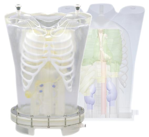
1. Radiology Phantoms PET/SPECT Thorax Phantom_PH-63
페이지 정보
작성일 22-07-22 14:03본문
Examination of myocardial density through SPECT imaging
-Verification of myocardial imaging with the use of various RI solution densities
-Ability to capture defects of the myocardial region
-Can reproduce image variations of the heart by injecting RI solutions in the liver, kidney and lungs
Examination of RI solution density for simulated tumors
-The simulated tumors can be inserted into lung, liver andbreast
-Tumors can be filled with FDG/RI solution into the spheresfor evaluation of density, size and placement
Training skills / Applications
- PET/SPECT
- Quality management of NM equipment
- Myocardial density with SPECT imaging
- RI solution density for tumor imaging
Case / Pathology
- Anatomy
Liver
Lung (right/left)
Kidney (right/left)
Hot spots (liver, lungs and breast)
* Hot spot for PET can be set in liver, lungs and breast.
Heart - Anatomical type:right ventricle, left ventricle and myocardium
Geometric type:left ventricle and myocardium
- 이전글X-Ray Training Phantom "PBU-POSE" 22.07.22
- 다음글Multi Energy CT Quality Assurance Phantom (TR-J) 22.07.21


